At Amazing Eye Care, our certified optometrist is skilled in diagnosing and treating glaucoma. With the use of advanced technology like the Optomap Daytona, we are able to detect early signs of glaucoma through thorough screening and annual scans.
Learn about the various tests eye doctors use to accurately diagnose glaucoma. A comprehensive evaluation includes assessing eye pressure, cornea thickness, and optic nerve health. This article explains the essential assessments and tests needed for a proper glaucoma diagnosis.
Risk Factor Assessment
Your eye doctor will begin the assessment by examining your risk factors for developing glaucoma, which will determine how often and how extensively further testing will be conducted. The evaluation includes a review of your family history and medical background to identify important risk factors such as:
- Age over 60
- Ethnic background, such as African or black Caribbean descent, Hispanic, or Asian
- Family history of glaucoma (e.g., a sibling or parent with glaucoma)
- History of eye conditions, injuries, or surgeries
- Prolonged corticosteroid use (eye drops, pills, inhalers, or creams)
- Chronic conditions affecting blood flow, such as migraines, diabetes, low blood pressure, or hypertension
- Current or former smoking status
For those who have undergone a comprehensive eye exam, additional factors considered include:
- Elevated eye pressure (above 21 mm Hg)
- Thin corneas (less than 500 um)
The type of eyesight can also impact the risk of developing different types of glaucoma. Farsightedness is associated with a higher risk of narrow-angle glaucoma, which progresses rapidly, while nearsightedness is linked to open-angle glaucoma, a slow-progressing type that may not have symptoms.
Common Glaucoma Tests
During a thorough eye examination, your eye doctor will always check for glaucoma, regardless of your risk factors. The important tonometry and ophthalmoscopy tests provide a starting point for future evaluations as you get older.
Tonometry
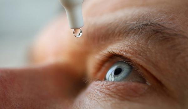
Tonometry is a procedure that measures the pressure inside your eye. Your eye doctor will use numbing eye drops and a tonometer to gently puff warm air onto your eye. While eye pressure can vary and may not always indicate glaucoma, it is still important information for your eye doctor to consider. Normal eye pressure usually falls between 12 and 22 mm Hg, with glaucoma often being diagnosed at pressures over 20 mm Hg. However, it is possible for individuals to have glaucoma even within the normal pressure range of 12 to 22 mm Hg.
Ophthalmoscopy
During your eye exam, your doctor will use eye drops to dilate your pupil and examine your optic nerve for shape and color. They will then use a small device with a light to magnify the optic nerve. Depending on the results, your doctor may recommend additional glaucoma tests.
Additional Glaucoma Tests
Perimetry
Perimetry is a test that maps your entire field of vision by having you signal when a moving light enters your peripheral vision while looking straight ahead. It is important to stay relaxed and provide accurate responses during the test. Your doctor may repeat the test to ensure consistency. If you have glaucoma, it is usually recommended to have an annual visual field test to monitor any changes in your vision.
Gonioscopy
This diagnostic exam evaluates the angle between your iris and cornea. After administering numbing eye drops, a handheld contact lens is delicately placed on the eye. Through a mirror on the contact lens, the doctor checks if the angle is closed and obstructed (which could indicate potential angle-closure or acute glaucoma) or wide and open (which may suggest potential open-angle or chronic glaucoma).
Pachymetry
Your eye doctor may use pachymetry as an additional diagnostic tool to measure the thickness of your cornea, the transparent front window of the eye. A pachymeter probe is gently placed on the cornea to obtain thickness measurements, which can enhance diagnostic accuracy as corneal thickness can affect eye pressure readings. Regular eye exams are crucial for early detection of glaucoma, especially if you have any risk factors.
Optic Nerve Examination
The optic nerve can be easily examined using an ophthalmoscope in a clinic. It exits through the back of the eye and is composed of over one million individual nerve fibers that originate in the retina. These fibers travel to different parts of the brain. When looking into the eye, the optic nerve is seen end on, and the nerve fibers can be faintly observed fanning out onto the retina.
In a normal state, the optic nerve head resembles a doughnut, with the outer ring made up of nerve tissue. The central nerve head contains a hole called the optic cup, which is the empty space left after the nerve fibers spread into the retina. In cases of glaucoma, the nerve fibers become damaged and deteriorate, resulting in a larger cup or hole in the doughnut shape.
A healthy optic nerve head typically has a thick outer ring of nerve tissue surrounding a small optic "cup" in the center. However, in cases of glaucomatous optic nerve damage, the outer ring becomes thin and the "cup" enlarges, indicating the loss of nerve fibers. While various diseases can affect the optic nerve, the distinct appearance caused by glaucoma allows ophthalmologists to identify its presence.